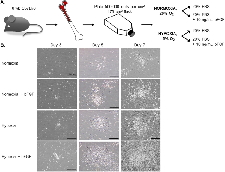
Figure 1.
MSC Culture Condition Development. (A) Schematic illustrating the workflow and conditions used to optimize the culture of mMSC. The fresh isolated bone marrow cells were divided into 4 groups: normoxic (20% O2) or hypoxic (5% O2) conditions +/−10 ng/mL of bFGF supplement. (B) The representative phase contrast images were acquired (10x) at different time points during the p0 to p1 growth phase: d3, d5, and d7 post-plating. (d14 images found in Fig. 2A). Scale bars = 500 μm.

