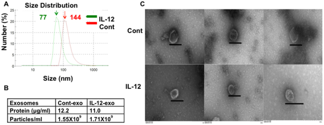
Figure 1.
Characterization of CTL-derived vesicles. Naïve CD8+ T cells purified from OT-I mice were stimulated with 2SI (cont) or 3SI (IL-12) for three days in vitro, and extracellular vesicles were purified from each supernatant. (A) Size distribution measured in Malvern Zetasizer Nano ZS90. (B) Concentration of protein and vesicle particles per mL of supernatant. (C) Transmission Electron Microscopy. Purified vesicles were observed under Zeiss EM10 transmission electron microscope. The graphs vary on amplification magnitude, and the bar in each graph indicates 200 nm. Each experiment was replicated at least three times with similar results.

