Figure 2.
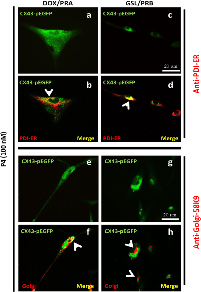
Cx43 forward trafficking from ER to Golgi is blocked in PRB expressing cells. Representative images of immunofluorescence on hTERT-HMA/B cells induced for PRA (left panel) or PRB (right panel) expression and stimulated with P4 (100 nM). Images show Cx43 in green fluorescence (a,c,e,g), Endoplasmic reticulum (ER) marker(PDI, b,d) and Golgi (G) marker (Golgi-58K9, f,h) in red colour, and co-localization of Cx43 and ER/G in yellow [Cx43 + ER (b,d) and Cx43 + G (f,h)]. PRB expressing cells show accumulation of Cx43 in ER (d, yellow) but not in Golgi (h, red) in the merged images. Scale bar = 20 μm.

