
Figure 4.
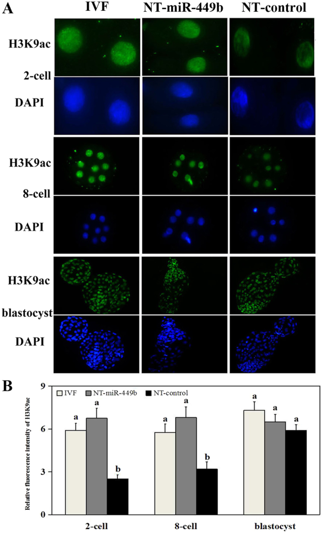
Immunofluorescence analysis of H3K9ac in various bovine embryos. (A) Global acetylation level of H3K9 (green) in embryos at the 2-cell, 8-cell, and blastocyst stages in IVF, NT-control, and NT-miR-449b groups. Each sample was counterstained with DAPI to visualize DNA (blue). Original magnification, 200×. (B) Relative fluorescence intensities of H3K9ac (using Image-Pro 6.0 software, and the values were showed as mean ± SEM). a,b Values with different superscripts within columns are significantly different from each other (P < 0.05).