Fig. 3.
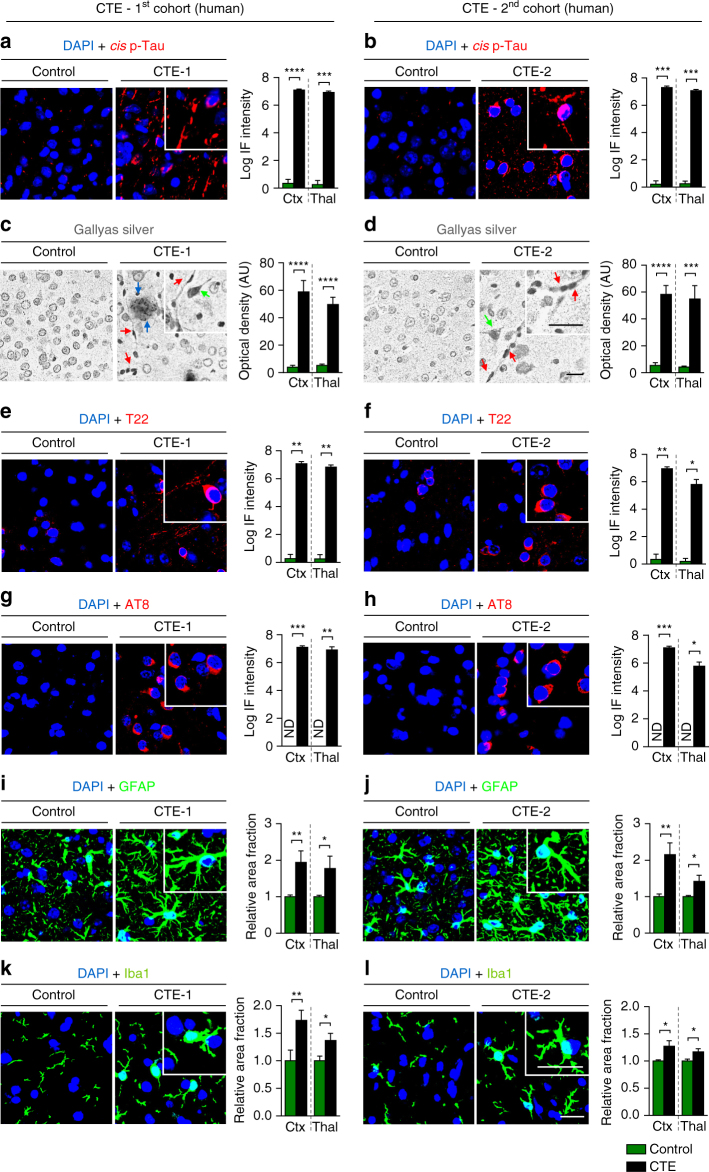
Cis P-tau is expressed in both the cortex and deep brain regions and correlates with various neuropathological features in CTE patients. CTE brain sections of the cortex and thalamus from eight collision sport athletes under ages of 75 from two independent sources and age-matched controls (Supplementary Table 3) were subjected to immunostaining or Gallyas silver staining, followed by confocal microscopy and light microscopy, respectively, to detect cis P-tau and various CTE pathologies. Cis P-tau staining a, b was correlated with the presence of axonal pathology, as detected by Gallyas silver staining c, d, tau oligomerization (T22) e, f, early tangles (AT8) g, h, astrogliosis (GFAP) i, j and microgliosis (Iba1) k, l in two different brain regions. Inset images are the high magnification image of selected area denoted by the white. Of note, there was not detectable signals when secondary antibodies were used alone (data not shown). Scale bar, 40 μm. Different types of pathologies in CTE brains revealed by Gallyas silver staining: senile plaque (blue arrow); neurofibrillary tangles (green arrow); axonal bulb (red arrow). Ctx, parasagittal cortex; Thal, thalamus; ND, not detectable; NS, not significant. Images were quantified and results are shown as means ± S.E.M. and p-values calculated using unpaired two-tailed parametric Student’s t-test. *p < 0.05, **p < 0.01, ***p < 0.001, ****p < 0.0001

