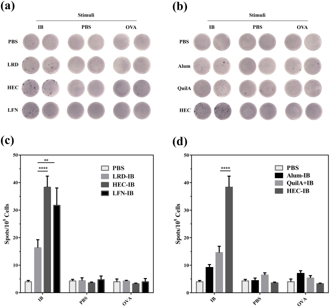
Figure 4.
Interferon-γ (IFN-γ) secretion by splenocytes from C57BL/6 J mice (n = 4/group) 49 days after vaccination with IB in combination with commercially available adjuvants (QuilA or Alum), nanoparticles (HEC, LRD or LFN) or PBS alone (control). Splenocytes (1.0 × 10⁵ cells/well) were harvested and co-cultured with IB (specific antigen), OVA (irrelevant antigen) or PBS (no antigen) for 40 h, then secretion of interferon-γ (IFN-γ) determined by ELISPOT assay. Photomicrographs show IFN-γ expressing splenocytes in response to (a) three hectorites and (b) best clay nanoparticle or commercial adjuvants; bar charts (c) and (d) illustrate the number of IFN-γ secreting spots in each vaccination group after stimulation with antigen (IB or OVA) or PBS. Data are expressed as mean ± S.E.M. (n = 4). *P < 0.05; **P < 0.01; ***P < 0.001; and ****P < 0.0001.

