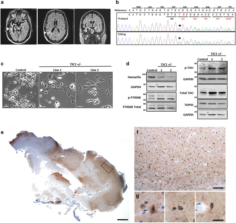
Fig. 1.
Proband clinical MRI, novel TSC1 mutation, cell model of TSC1/hamartin haploinsufficiency, and neuropathology of a TSC1 mutation carrier. a Magnetic resonance imaging (MRI) FLAIR sequences revealed severe atrophy of the right anterior temporal lobe, particularly the entorhinal/perirhinal cortices and inferior temporal gyrus, as well as the hippocampus and amygdala. There was also involvement of the left anterior/mesial temporal lobe and the bilateral frontal and right parietal lobes. T2/FLAIR hyperintensities are seen within the subcortical white matter, some of which extended to the superolateral aspects of the lateral ventricles (arrowheads). These lesions can be seen in tuberous sclerosis or focal cortical dysplasias as a transmantle sign. b Sanger sequencing confirmed a novel frameshift variant in the TSC1 gene (NM_000368.4: c.62_63insTG: p. Arg22CysfsTer5) that segregated with the proband and affected sibling. c Representative micrographs of the enlarged cell body in the undifferentiated TSC1 heterozygous mutant SH-SY5Y lines. Scale bars represent 50 microns. d Representative western blots showing decreased TSC1/hamartin and increased phopho-P70S6Kthr389, total tau, and phospho-tau levels in retinoic acid differentiated TSC1 +/− SH-SY5Y cells. TDP-43 levels are unchanged. e–g Fragment of frontal cortex resection from the proband’s sibling was received and stained for hyperphosphorylated tau, revealing patchy staining in neurons, glia, and surrounding neuropil. Scale bars represent 1000 (e), 250 (f), and 25 (g) microns

