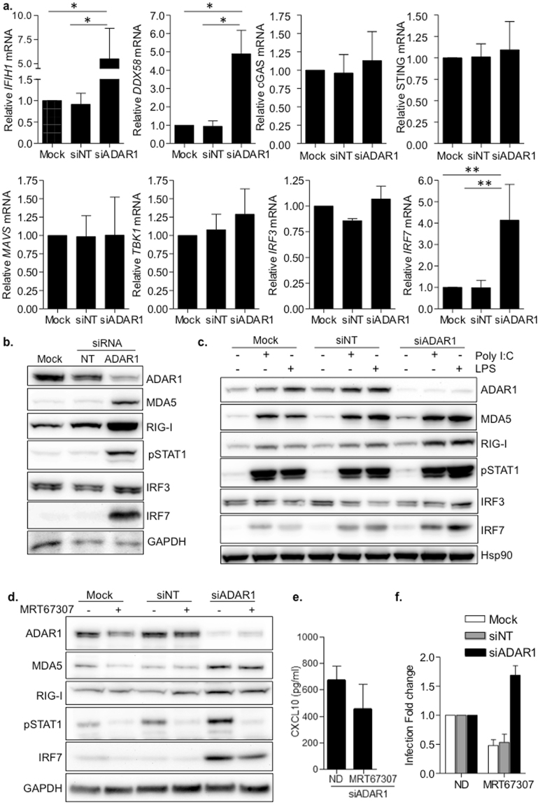
Figure 4.
ADAR1 specifically regulates the RLRs-MAVS signaling pathway. (a) Relative mRNA expression of RLRs (IFIH1 and DDX58), DNA sensors (cGAS and STING), downstream signaling proteins (MAVS, TBK1) and transcription factors (IRF3 and IRF7) in ADAR1 knockdown macrophages. Data represents mean ± SD of at least 4 different donors and is normalized to Mock-transfected macrophages. *p < 0.05; **p < 0.005. (b) Protein expression of RLRs and related proteins in macrophages, showing overexpression of MDA5, RIG-I, pSTAT1 and IRF7 consequence of ADAR1 inhibition. A representative donor is shown. (c) Western blot showing protein expression pattern in transfected macrophages, stimulated or not with LPS (100 ng/ml) or Poly I:C (10 µg/ml) to resemble canonical TLR-mediated activation of type I IFN response. A representative donor is shown. (d) Protein expression in ADAR knockdown macrophages treated with the TBK1 inhibitor MRT67307 (5 µM). Blocking TBK1 function partially restores protein expression phenotype observed in Mock- or siNT-transfected macrophages. A representative donor is shown. (e) CXCL10 protein expression in the supernatant in ADAR1 knockdown macrophages, treated or not with MRT67307 (5 µM). CXCL10 protein in the culture supernatants was measured by ELISA. Data represents mean ± SD of 3 different donors. (f) Change in HIV-1 replication of macrophages treated with the TBK1 inhibitor MRT67307 (5 µM). Fold change of HIV-1 infection in macrophages treated or not with MRT67307. Infection is normalized to the corresponding untreated condition. Data represents mean ± SD of 3 different donors performed in triplicate. *p < 0.05; **p < 0.005. The figure shows the cropped gels/blots obtained by each protein evaluation. Full-length blots of each tested protein are included in supplementary material.

