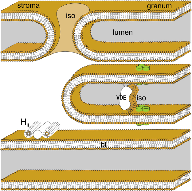
Figure 6.
Schematic representation of thylakoid membranes of vascular plants – showing the tentative assignments of the lipid phases detected by 31P-NMR. Lipid phases: the basic bilayer (bl) structure, the non-bilayer, isotropic phases (iso) associated with the fusion of granum and stroma thylakoid membranes and with the lumenal lipocalin proteins,VDE and CHL, as well as the HII phase in the stroma – possibly also associated with membrane-associated proteins and loosely attached to the membrane. The figure is not to scale; for simplicity, CURT1 proteins, which are enriched in the end-membranes of thylakoids and maintain the extreme curvature of membranes at the margins75, are omitted. Membrane-intrinsic proteins are symbolized by trimeric LHCIIs (green bars).

