
Figure 5.
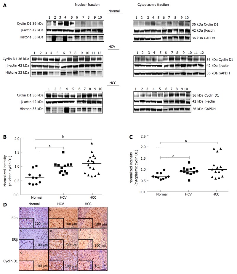
Expression of cyclin D1 in nuclear and cytoplasmic tissue lysates from normal and diseased subjects. A: Lysates prepared from nuclear and cytoplasmic fractions of liver tissues from normal, HCV and HCC subjects were subjected to Western blotting and probed with antibodies against cyclin D1, β-actin, histones (nuclear lysates only) and GAPDH (cytoplasmic lysates only). Representative blots for each group are depicted. B and C: The bands corresponding to cyclin D1 and β-actin were quantified by densitometric analyses using ImageJ. Expression of cyclin D1 was normalized to the expression of β-actin in nuclear (B) and cytoplasmic (C) lysates and plotted. aP < 0.05 or bP < 0.001 was considered significant. D: Immunohistochemical staining of ERα, ERβ and cyclin D1 in normal (a, d, g), HCV-related cirrhosis (b, e, h) and HCV-related HCC (c, f, i) is depicted. A magnified region of the stained tissue section is depicted as an inset in each corresponding image. Scale bar = 100 μm. HCV: Hepatitis C virus; HCC: Hepatocellular carcinoma.