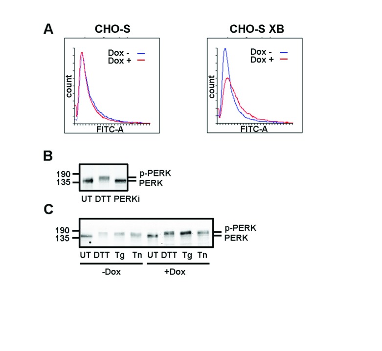
Figure 4. PERK activation is unchanged by overexpression of XBP1S.
( A) Flow cytometry analysis of fluorescence from CHO-S and CHO-S XB cells stained with fluorescent ER Tracker dye. Samples were either treated with doxycycline (Dox) for 3 days (red) or left untreated (blue). ( B) Western blot of lysates from CHO-S XB cells that were either untreated (UT), treated with a reducing agent (DTT) or with an inhibitor of PERK kinase activity (PERKi). Blots were probed with anti-PERK to display the extent of PERK phosphorylation. ( C) Anti-PERK western blot of CHO-S XB cells induced with Dox and subsequently treated with DTT, thapsigargin (Tg) or tunicamycin (Tn). Experiment ( A, B and C) were performed twice.

