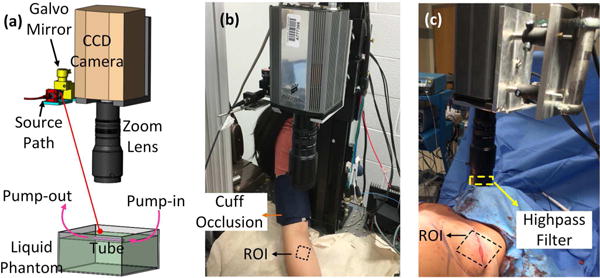
Fig. 1.

The nc_scDCT instrument and experimental setups for 3D flow imaging. (a) In the first heterogeneous phantom, a transparent cylindrical tube, connected with a peristaltic pump, was placed at ~5 mm below the liquid phantom surface to create high flow contrasts against the background. In the second heterogeneous phantom, two spherical solid phantoms were placed at ~5 mm below the liquid phantom surface at different locations to create low flow contrasts. (b) Blood flow changes in a healthy human forearm induced by the arterial cuff-occlusion on subject’s upper arm were continuously imaged by nc_scDCT. (c) A mastectomy skin flap imaged by nc_scDCT intraoperatively. An 800-nm high-pass filter minimizes the influence of ambient light in the operating room after moving away the lamp beam from the breast. For laboratorial experiments, the room was kept dark.
