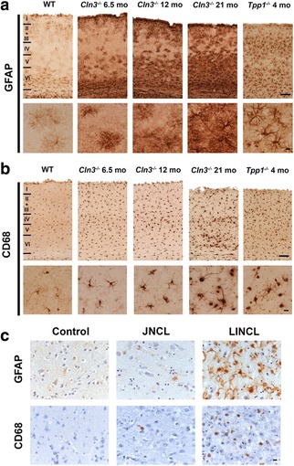
Fig. 1.

Attenuated glial responses in Cln3 −/− mouse tissue and in human JNCL. Cortical sections from wild type (WT), Cln3 −/− and Tpp-1 −/− mice (a, b) or from LINCL and JNCL human cases (c) were immunostained with Glial Fibrillary Acid Protein (GFAP) or Cluster of Differentiation 68 (CD68) to investigate the level of reactive astrocytosis or microglial activation, respectively. Compared to the very low level of glial activation present in WT mice (shown at 6.5 months of age, but changes very little over time), marked astrocytosis (a) and microglial activation (b) was apparent in severely affected 4-month-old Tpp-1 −/− mouse sections, with hypertrophied astrocytes with intense GFAP immunostaining and thickened processes, and morphologically transformed microglia with intense CD68 immunostaining being observed. a In contrast, astrocytosis appeared different in nature in the Cln3 −/− cortex, and although many astrocytes displayed intense GFAP immunoreactivity, especially in the deeper laminae, most these astrocytes still retained many thin processes reminiscent of protoplasmic astrocytes. Even at the end stages of the disease (21-month-old Cln3 −/− mice) very few astrocytes became fully hypertrophied. b A similar attenuated morphological transformation of microglial cells was also apparent in the cortex of Cln3 −/− mice. Although microglia became more intensely CD68 immunoreactive and more swollen with increased age, many of these cells retaining long branched processes, even in 21-month-old Cln3 −/− mice. (c) Reactive astrocytosis was observed in both human INCL and JNCL (c), but to very different extents. In LINCL cases astrocytes were intensely immunostained, with hypertrophied cell bodies and numerous thickened processes. In JNCL cases GFAP immunostaining was paler and far fewer, less hypertrophied astrocytes with thinner processes were evident. Only a few CD68 positive microglia were observed in JNCL cases (c), compared to the relative abundance of activated CD68 immunoreactive microglia activation present in the cortex of LINCL cases. Scale bars = 120 μm (a, b) 20 μm in higher magnification views; 50 μm (c). Roman numerals in a, b indicate cortical laminar boundaries
