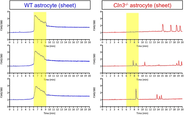
Fig. 10.

Cln3 −/− Astrocytes Show Altered Calcium Signalling. Recordings of Fura-2 fluorescence were made from high density, sheet forming cultures of wild type (WT) and Cln3-deficient (Cln3 −/−) astrocytes grown under basal conditions over a period of 30-45 min, from which the first 20 min are shown. The figure illustrates changes in [Ca2+]I in three randomly selected WT and Cln3 −/− astrocytes. In response to treatment with 100 μM ATP, a propagating [Ca2+]I wave was generated by WT astrocytes (marked with yellow bar). This synchronized [Ca2+]I wave had a large amplitude, and a prolonged plateau persisting for several minutes after initiation. The Cln3 −/− astrocytes did not exhibit any propagating calcium waves, instead, Cln3 −/− astrocytes had non-synchronized, spontaneous [Ca2+]I elevations. Data is presented as 340 nm/380 nm ratio, which directly correlates with the change in intracellular free Ca2+ levels
