
Fig. 6.
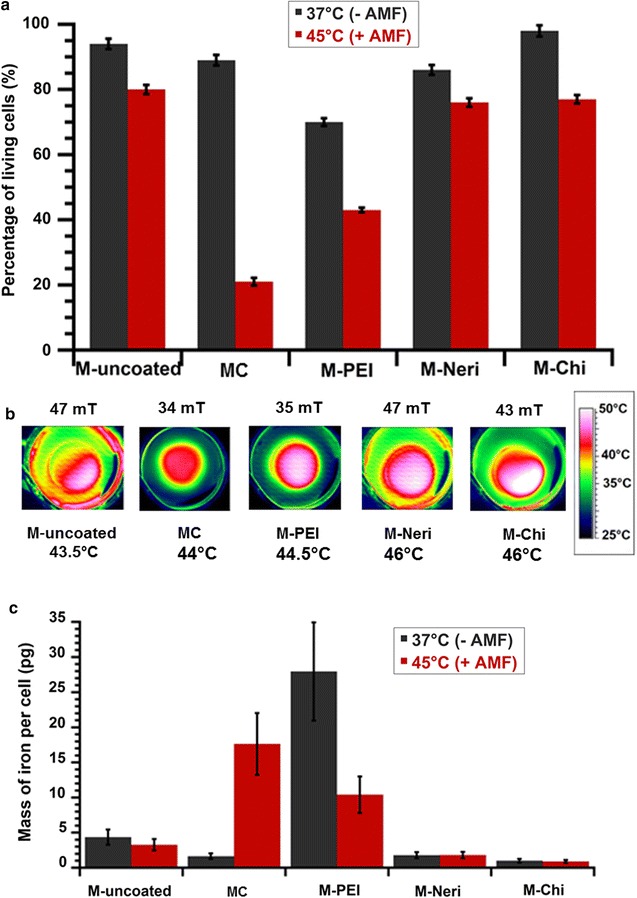
a Percentage of living GL-261 cells (%) when these cells are brought into contact with 1 mg/mL of MC, M-uncoated, M-Neri, M-Chi, and M-PEI, and either maintained at 37 °C during 30 min without AMF treatment, black columns: 37 °C (−AMF), or exposed during 30 min to an AMF of frequency 198 kHz and strength varied between 34 and 47 mT to maintain temperature at between 43 and 46 °C during 30 min, red columns: 45 °C (+AMF). b Spatial temperature distribution, measured with an infra-red camera, of GL-261 cells brought into contact with 1 mg/mL of MC, M-uncoated, M-Neri, M-Chi or M-PEI and exposed during 30 min to the same AMF as in a. c Quantity of iron coming from magnetosomes, which is internalized in each GL-261 cell when GL-261 cells are brought into contact with 1 mg/mL of MC, M-uncoated, M-Neri, M-Chi, or M-PEI, and exposed during 30 min to the same AMF as in a