Fig. 1.

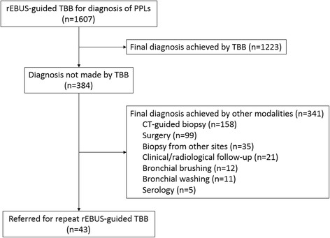
Study flow diagram. CT, computed tomography; rEBUS, radial-probe endobronchial ultrasound; PPL, peripheral pulmonary lesion; TBB, transbronchial biopsy

Study flow diagram. CT, computed tomography; rEBUS, radial-probe endobronchial ultrasound; PPL, peripheral pulmonary lesion; TBB, transbronchial biopsy