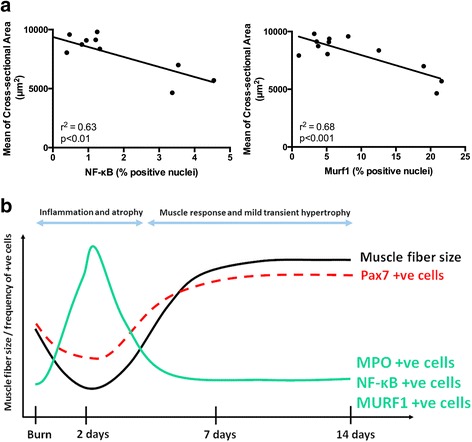
Fig. 6.

The abundance of NF-κB- and Murf1-positive nuclei in muscle tissue correlates with muscle atrophy. a Correlation plots between the percentage of NF-κB-positive nuclei and the mean of myofiber cross-sectional area (r2 = 0.63, P < 0.01), and the percentage Murf1-positive nuclei and the mean of myofiber cross-sectional area (r2 = 0.68, P < 0.001). Correlation analysis was performed using two-tailed Pearson correlation. The analysis includes mice from all treatment groups (sham and 2 days, 7 days, and 14 days post-burn). b Cellular and molecular cascades associated with muscle atrophy during the acute burn response and subsequent muscle regrowth. MPO myeloperoxidase
