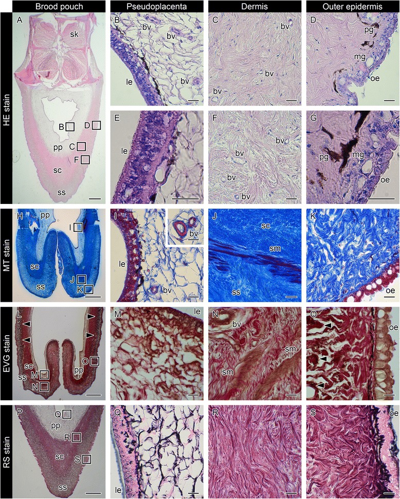
Fig. 1.

Histology of the brood pouch in mature pot-bellied seahorse Hippocampus abdominalis. Hematoxylin and eosin (HE) stain of cross-sections of the brood pouch (a–g), Masson’s trichrome (MT) stain of a cross-section around the entrance of the pouch (h–k), Elastica van Gieson (EVG) stain of a cross-section around the entrance of the pouch (l–o), and reticulin silver (RS) stain of a cross-section of the brood pouch (p–s). The dorsal side is at the top and the ventral side is at the bottom in a, h, l, and p. The lettered boxes in a, h, l and p indicate sites of high magnification of pseudoplacenta (b, i, m, q), dermis (c, f, j, n, r), and outer epidermis (d, k, o, s). High-magnification views of pseudoplacenta (e) and outer epidermis (g). The inset panel in (i) shows the large blood vessels found in pseudoplacenta. Arrowheads indicate black signals after EVG staining. Scale bars: a, h, l, p = 1 mm; b–g, i–k, m–o, and q–s = 20 μm. bv, blood vessel; le, luminal epithelium; mg, mucous granule; oe, outer epithelium; pg, pigment cell; pp, pseudoplacenta; sc, stratum compactum; sk, skeletal muscle; sm, smooth muscle; ss, stratum spongiosum. These photographs were taken using two individual male seahorses; one specimen for a, h, p, and another for l, due to the lack of black staining in the first specimen
