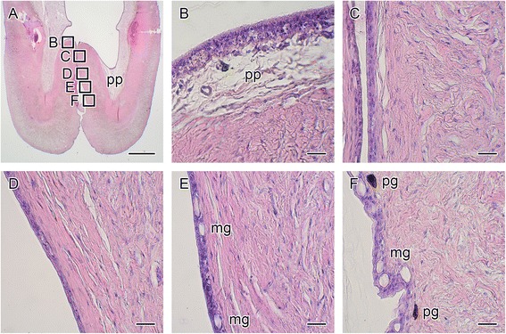
Fig. 2.

Hematoxylin and eosin staining around the entrance of the seahorse brood pouch. A cross-section of the brood pouch around the entrance is shown in a. The dorsal side is at the top and the ventral side is at the bottom. The lettered boxes in a indicate sites of high-magnification views of the luminal epithelium (b) to the outer epithelium (c–f). mg, mucous granule; pg, pigment cell; pp, pseudoplacenta. Scale bars: a = 1 mm; b–f = 20 μm. The same individual (also depicted in Fig. 1 a) is shown in all photographs
