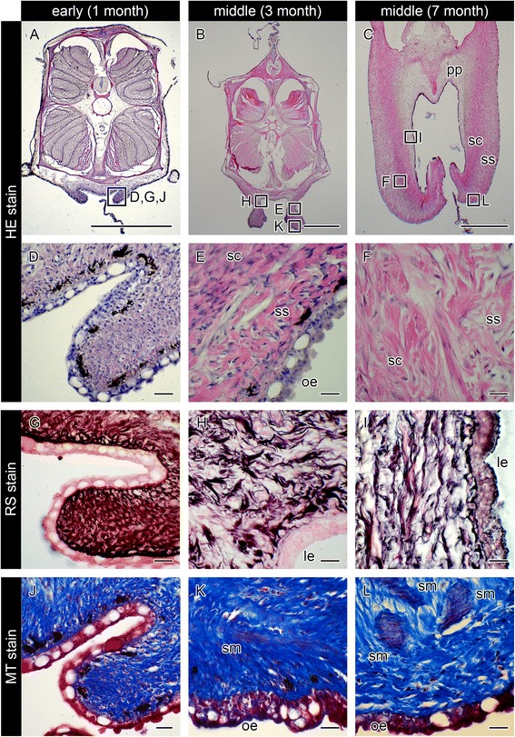
Fig. 4.

Histological observations of seahorse brood pouch formation. Cross-sections of the brood pouch were observed by various staining methods: hematoxylin and eosin (HE) stain (a–f), reticulin silver (RS) stain (g–i), and Masson’s trichrome (MT) stain (j–l). These photographs show three individual males: one specimen for a, d, g, j, at 20–30 days after birth, showing the Y-shaped seam line (the same individual as in Fig. 3 d); one specimen for b, e, h, k, at three months after birth; and one specimen for c, f, i, l, at seven months after birth. The lettered boxes in a–c indicate the corresponding sites of high-magnification views shown in d–l. Scale bars: a–c = 1 mm; d–l = 20 μm. le, luminal epithelium; oe, outer epithelium; pp, pseudoplacenta; sc, stratum compactum; sm, smooth muscle; ss, stratum spongiosum
