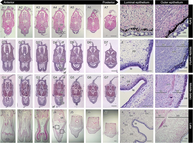
Fig. 5.

Hematoxylin and eosin stain of serial sections throughout development of the seahorse brood pouch. Serial sections of the body: at the early stage of pouch development showing the I-shaped seam line (a1–7; the same individual as in Fig. 3e); in the early period of the middle stage (d1–7), and the latter period of the middle stage (g1–7); and at the late stage (j1–7). The section views are from the anterior (1) to posterior (7). The lettered boxes in a4, d4, g4, and j4 indicate sites of high magnification of the luminal epithelium (b, e, h, k) and outer epithelium (c, f, i, l). af, anal fin; bv, blood vessel; df, dorsal fin; le, luminal epithelium; oe, outer epithelium; pp, pseudoplacenta; sc, stratum compactum; sk, skeletal muscle; sm, smooth muscle; ss, stratum spongiosum. Scale bars: a, d, g, j = 1 mm; b, c, e, f, h, i, k, l = 100 μm. These photographs were taken using four individuals: one specimen for a–c, one for d–f, one for g–i, and one for j–l
