
Fig. 1.
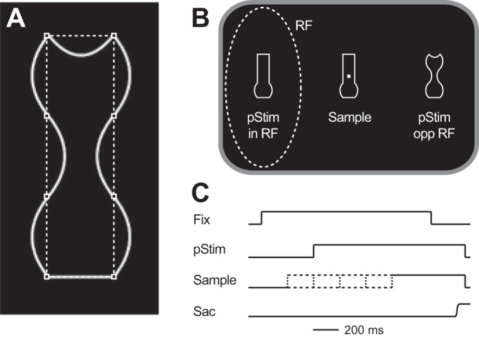
Behavioral task. A: each object was made up of 8 solid white lines that connected 8 imaginary points (squares). Each line could be straight, concave, or convex, and each object was vertically symmetrical. B: stimulus arrangement. On each trial a sample stimulus was presented around a fixation point and 2 stimuli were presented in the visual periphery (pStim), with 1 placed in the neuron’s response field (RF) and the other diametrically opposite. One of the peripheral stimuli matched the sample (match); the other did not (nonmatch). Stimuli were chosen from a set of 5 that were new in each session. C: time course of a trial. Each trial began (Fix) after the animal had fixated a central fixation point for 500–1,000 ms. After this the peripheral stimuli (pStim) appeared 400 ms later and remained on until the end of the trial. The sample could appear 200 ms before the peripheral stimuli, at the same time as the peripheral stimuli, or 200, 400, or 600 ms after the peripheral stimuli (indicated by dashed lines). We refer to these as having stimulus onset asynchronies (SOAs) of −200, 0, 200, 400, or 600 ms respectively. The animal had to make a saccade (Sac) to the match to obtain a reward.