Fig. 8.
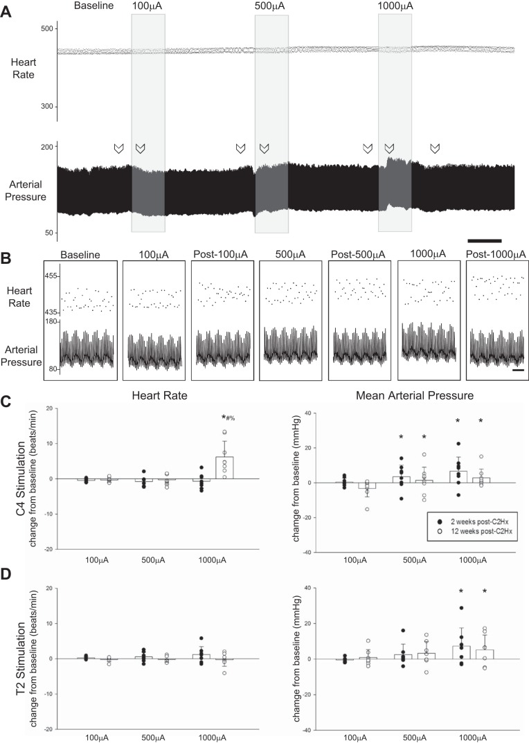
Impact of HF-ES on cardiovascular parameters. A: representative traces depict instantaneous heart rate (HR; beats/min) and mean arterial pressure (MAP; mmHg) from a rat that received stimulation to the C4 spinal cord 2 wk postinjury. B: expanded traces depict instantaneous HR and MAP at baseline as well as during and after each bout of stimulation. Arrows in A indicate locations from which the expanded traces shown in B were obtained. Scale bars represent 1 min (A) and 1 s (B). C and D: stimulation-induced changes in HR and MAP for rats receiving HF-ES at either the C4 (C) or the T2 (D) spinal level. A significant increase in HR was observed following C4 stimulation in rats that were 12 wk postinjury. MAP was significantly increased relative to baseline during stimulation at both 500 (#P < 0.05) and 1,000 µA (*P < 0.05). Following T2 stimulation, MAP was only increased with stimulation at 1,000 µA (*P < 0.05). In all groups, HR and MAP returned to baseline following cessation of stimulation. *P < 0.05, different from 100 µA. #P < 0.05, different from 500 µA. %P < 0.05, different from 2 wk. Data were evaluated using 2-way repeated-measures ANOVA with Holm-Sidak post hoc tests for individual comparisons (groups: 2-wk C4, n = 8; 12-wk C4, n = 8; 2-wk T2, n = 7; 12-wk T2, n = 7).

