
Fig. 3.
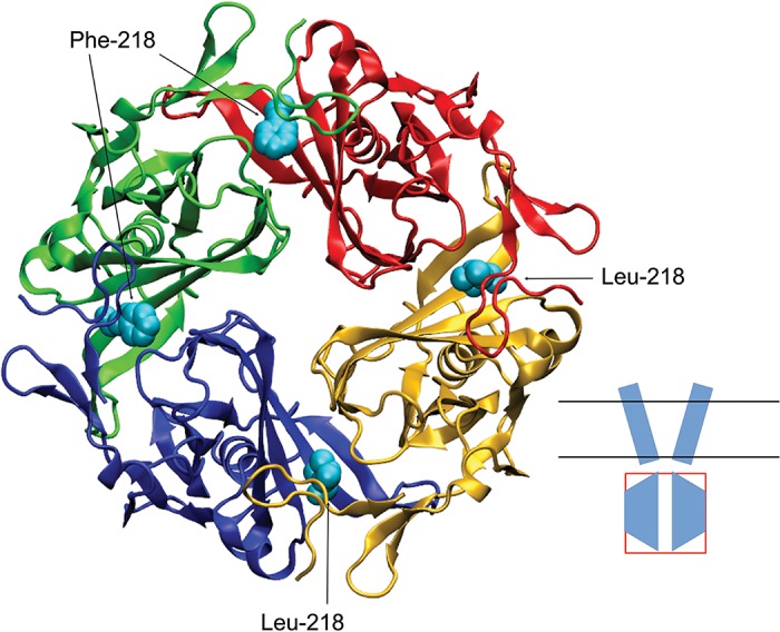
Three-dimensional homology model of the intracellular domain of Kir4.1 WT/L218F channel. Model indicates the structural difference of a larger phenylalanine at site 218 in the red and green subunits compared with that of a leucine, shown in the blue and yellow subunits. Inset shows a cartoon of the channel in which the intracellular domain is boxed.