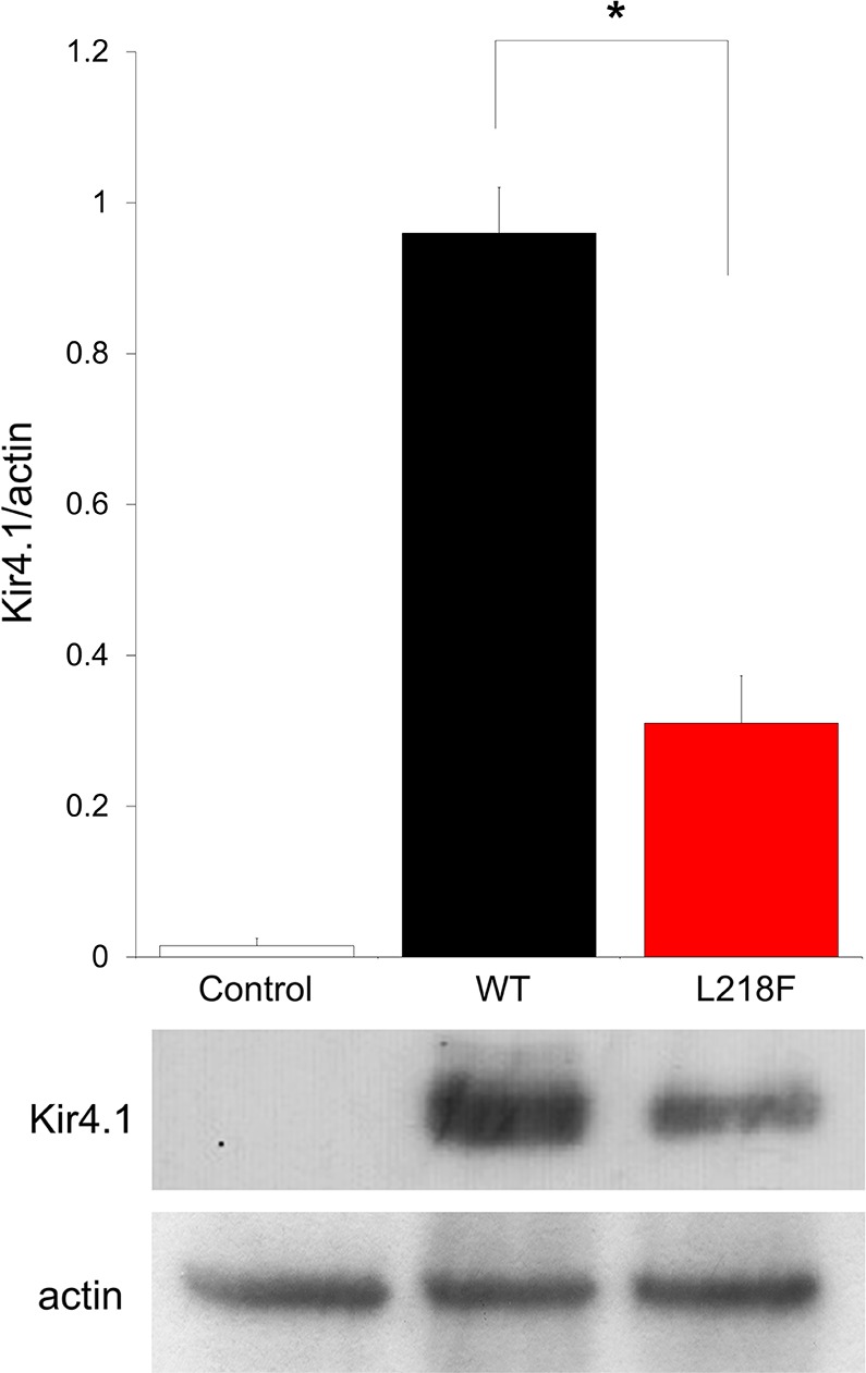
Fig. 4.

Recombinant Kir4.1 expression in Xenopus oocytes. Shown is densitometric analysis (top) of recombinant Kir4.1 bands (bottom) derived from total membrane protein extracts from mock-injected oocytes (left), WT cRNA-injected oocytes (middle), or L218F cRNA-injected oocytes (right), detected with anti-Kir4.1 antibody and normalized to the corresponding actin value, which was used as loading control. One representative immunoblot of oocyte lysates of 3 is shown. Data are means ± SE of 3 independent experiments (*P < 0.01 vs. WT).
