Fig. 6.
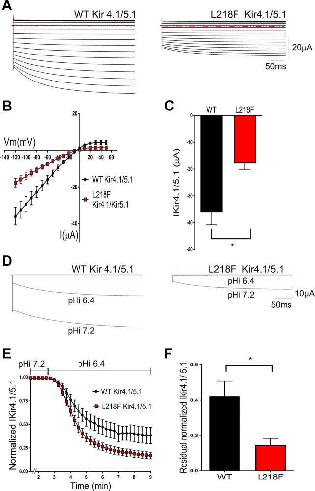
Effects of L218F mutation on heteromeric Kir4.1/Kir5.1 channel function. A: representative current families from oocytes expressing WT Kir4.1/Kir5.1 and L218F Kir4.1/Kir5.1 channels. Currents were evoked by voltage commands as described in Fig. 5, and all displayed the time-dependent “relaxation” typical of Kir4.1/Kir5.1 current. Horizontal red dashed lines indicate 0 current level. B: average steady-state I-V relationships for WT Kir41/5.1 and L218F Kir4.1/5.1. The current amplitudes measured from oocytes injected with mutant cRNA were smaller than those measured from oocytes injected with WT cRNA. C: bar graph showing that at −120 mV, the mean L218F Kir4.1/Kir5.1 current is significantly smaller than the mean WT Kir4.1/Kir5.1 current (n = 8; *P < 0.005). I-V data and bar graphs are representative of independent experiments performed using 2 different batches of oocytes (n = 8). These results indicate that L218F mutation reduced heteromeric current amplitudes. D–F: effect of L218F mutation on the pH sensitivity of Kir4.1/Kir5.1 channels. D: representative currents recorded from oocytes expressing WT Kir4.1/5.1 and L218 Kir4.1/5.1 cRNA. Currents were recorded during the perfusion of a membrane-permeable potassium acetate buffer that reduced the oocyte pHi from 7.2 to 6.4 and at a test potential of −120 mV. E: heteromeric current inhibition with time. WT cRNA-injected oocytes had a time course curve significantly different from that of the mutant cRNA-injected oocytes (n = 10; P < 0.0001). Current amplitudes, recorded at a test potential of −120 mV, were normalized to the peak current before the pHi was changed. The 2 curves were fitted with the equation Y = plateau + (Y0 − plateau)·exp [−K·(X − X0)]. F: bar graph showing residual current after 10 min of potassium acetate buffer perfusion. Residual L218F Kir4.1/Kir5.1 current was significantly less than that of the WT (n = 9; *P = 0.01). Results are from 3 different batches of oocytes.

