
Figure 5.
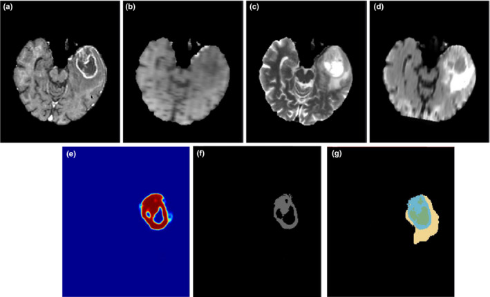
Illustration of the enhanced tumor segmentation using our proposed HNN method on one HGG patient from BRATS 2013 datasets. The four images in the first row shows the MRI modalities. (a) T1c, (b) T1, (c) T2, (d) Flair. Images in the bottom row from left to right are (e) prediction map, (f) final binary segmentation after optimal thresholding on prediction map, and (g) ground truth where light blue color represents enhanced tumor. [Color figure can be viewed at wileyonlinelibrary.com]