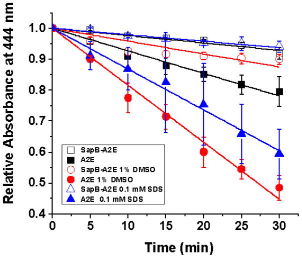
Figure 5.
Effect of 450–460 nm blue light on 40 μM A2E, free or bound to SapB (80 μM) followed over 30 min at 444 nm in 50 mM phosphate buffer with either 1 % methanol (not indicated), 0.1 mM SDS (open and filled triangles) or 1 % DMSO (open and filled circles). Experimental slopes are as follow: A2E (0.0068 SEM ±0.0003, R2 =0.984); SapB-A2E (0.0026 SEM ±0.0002, R2 =0.952); A2E 1% DMSO (0.0170 SEM ±0.0009, R2 =0.982); SapB-A2E 1 % DMSO (0.0030 SEM ±0.0006, R2 =0.784); A2E 0.1 mM SDS (0.0170 SEM ±0.0009, R2 =0.982); SapB-A2E 0.1 mM SDS (0.0030 SEM ±0.0006, R2 =0.784).

