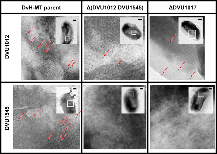
FIG 5 .
Electron microscopy images of DVU1012 (a) and DVU1545 (b) antibody-treated DvH-MT cells. Antibody binding of representative cells of the DvH-MT parental strain JWT700, the putative biofilm structural protein Δ(DVU1012 DVU1545) double mutant, and the ΔDVU1017 ABC transporter mutant. Binding is depicted by black spots of 10-nm-diameter gold particles, and examples are highlighted by red arrows. Images are enlargements of the white boxed region from the inset. Scale bars in the insets represent 200 nm.

