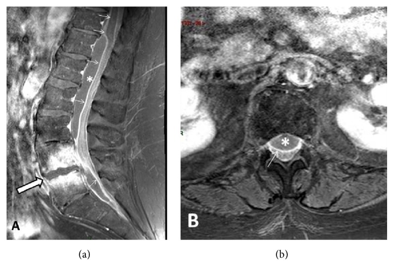
Figure 1.

Lumbar spine MR work-up at admission. (a) Midsagittal contrast-enhanced (CE) T1-weighted view with Fat Suppression (FS) option. Spondylodiscitis at L4-L5 level (arrow) with necroticocystic intersomatic abscess and strong enhancement of the adjacent bone marrow. Epidural abscess extending from T11/T12 to S1/S2 was well seen (asterisk) together with intense enhancement of the dura (arrows). (b) Axial–transverse CE T1-weighted view through the L1 level. Anterior epidural abscess is well seen (asterisk) impinging on posteriorly displaced thecal sac. Note intense enhancement of the dura (arrow).
