
Figure 7. Rapidly-adapting DRG neurons show identical frequency filtering to mouse Piezo2 in HEK293t cells.
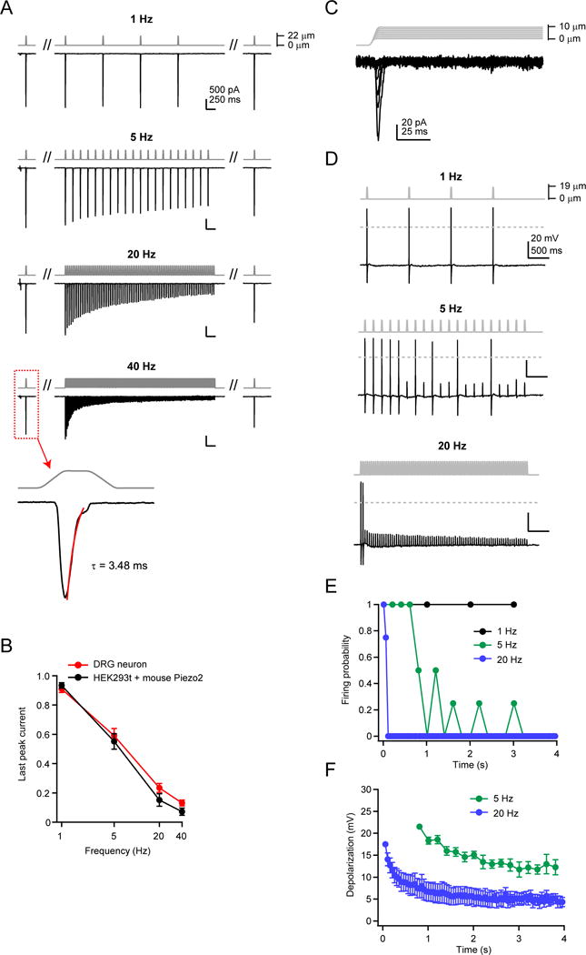
(A) Stimulus protocol and representative currents from a whole-cell recording from a single mouse DRG neuron, elicited by a 20 ms square pulse poke stimulus at 1, 5, 20, and 40 Hz. All four frequencies were tested on the same cell. The cell displayed rapidly-adapting mechanosensitive currents (inset, 40 Hz), with an inactivation time course of τ <10 ms (Ranade et al., 2014b). Holding potential was −80 mV. (B) Amplitude of the last peak current, normalized to ‘step1’, for rapidly-adapting DRG neurons (red, N = 7) and HEK293t cells transiently transfected with wild-type mouse Piezo2 (black, N = 10). (C) Stimulus protocol (gray) and representative current (black) from a rapidly-adapting DRG neuron in voltage clamp. (D) Stimulus protocol and action potentials elicited from the same neuron as in (C), in current clamp. Dashed line is 0 mV. (E) Firing probability for DRG neurons in response to 1, 5, and 20 Hz mechanical stimulation. Action potentials were counted for all responses crossing 0 mV. N = 4 neurons; all frequencies tested on each neuron. (F) Amplitude of subthreshold responses to 5 and 20 Hz from stimuli that failed to evoke action potentials in (E). All data are mean ± s.e.m.