Figure 2.
Human Platelet Accrual on Plasma VWFHA1 in Flow
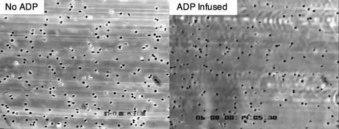
(A) Main chain schematic of the human von Willebrand factor (VWF)-A1 domain depicting residues that differ from its murine counterpart. Colored spheres denote the following: black are residues with buried side chains, green have partially buried side chains, red have exposed side chains on the front and upper surfaces, and gold have exposed side chains on the lower and back surfaces. Circled in blue is residue 1326, which plays a key role in permitting interactions with the GPIbα on human and mouse platelets (29). Accumulation of platelets on (B) surface-immobilized human plasma VWF or (C) murine plasma VWFHA1 is shown. Citrated whole blood from neonatal patients or healthy adults was perfused over the reactive substrate for 3 min (wall shear rate of 1,600 s−1) before assessing the number of interacting platelets. (D) Percentage of translocating platelets that became firmly adherent in response to infusion of buffer containing adenosine diphosphate (20 μM). The human GPIbα function blocking monoclonal antibody (mAb) 6D1 (10 μg/ml) or the αIIbβ3 inhibitor abciximab (10 μg/ml) was added to blood 10 min before use. See Supplemental Video 1. Data represent the mean ± SEM (n = 10 individuals per age group). ***p < 0.001 for antibody treatment versus no treatment according to Mann-Whitney U test. CHD = congenital heart disease.
P2Y Signaling Axis Is Robust in Neonatal Platelets and Can Support Integrin-Dependent Firm Adhesion to Surface-Adherent VWF in Response to ADP
Citrated whole blood from a neonatal cardiac patient was infused over surface-immobilized plasma von Willebrand factor (VWF)HA1 for 3 min (wall shear rate of 1,600 s−1), followed by the addition of platelet buffer with or without adenosine diphosphate (ADP) (20 mmol/L) for 1 min. ADP is seen to trigger firm adhesion, which prevents the further forward motion of translocating platelets.


