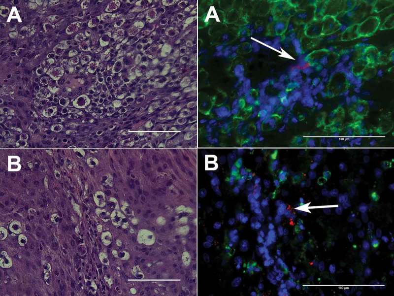
Figure 3.

Representative images of P. gingivalis antigen in the metrial triangle of SD rats in association with uNK cells (A) or CD68+ decidual macrophages (B). P. gingivalis (red) was detected with a rabbit polyclonal antibody to whole cell W83 [6,77], uNK cells were identified with mouse monoclonal antibody clone ANK61 to NK cell activation structure, decidual macrophages were labeled with anti-CD68 mouse clone ED1antibody (Abcam®, Cambridge, MA), and nuclei (blue) were stained with DAPI (results have not been published elsewhere).
