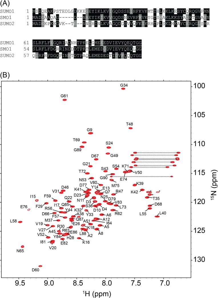
Fig 1. Sequence and structural examination of SMO-1.
(A) Multiple sequence alignment of SMO-1 with its homologues SUMO1 and SUMO2. Identical residues across the homologues are boxed in black while similar residues are boxed in grey. (B) 15N-HSQC of SMO-1 protein is shown with backbone amide peaks labelled by residue numbers. The side chain amides are connected by dashed lines. The folded peaks of arginine side chains are marked by an asterisk.

