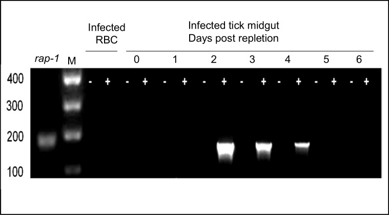
Fig 1. RT-PCR analysis of B. bovis hap2.
Analysis was performed in B. bovis from blood from an acutely infected animal, and in dissected tick midguts of female ticks that fed on animals acutely infected with B. bovis. Tick samples were obtained at 0–6 days post-repletion. Amplifications were performed on samples with (+) or without (-) the addition of reverse transcriptase. RBC: red blood cells. rap1: positive control. Molecular size markers in base pairs are indicated on the left side.

