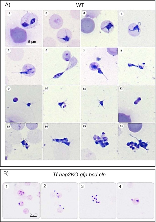
Fig 4. Photomicrograph of Giemsa-stained B. bovis smears.
A. Morphological changes of B. bovis wild type parasites developing in induced in vitro cultures, (1–4) lntraerythrocytic parasites, develop ray bodies (Strahlenkorper) whilst still intracellular. (5–7) Strahlenkorper egressing from erythrocytes. (8–11) Polymorphic multi-nucleated population of Strahlenkorper with long and short spikes. (12, 13) Two strahlenkorper adhering to each other along cell membranes. (14–16) Large aggregates of adhering multi-nucleated Strahlenkorper, occurring 20 h after induction. B. B. bovis Tf-hap2KO-gfp-bsd-cln parasites with induction medium (XA) at 26°C (1–4). Bars, 5 μm.

