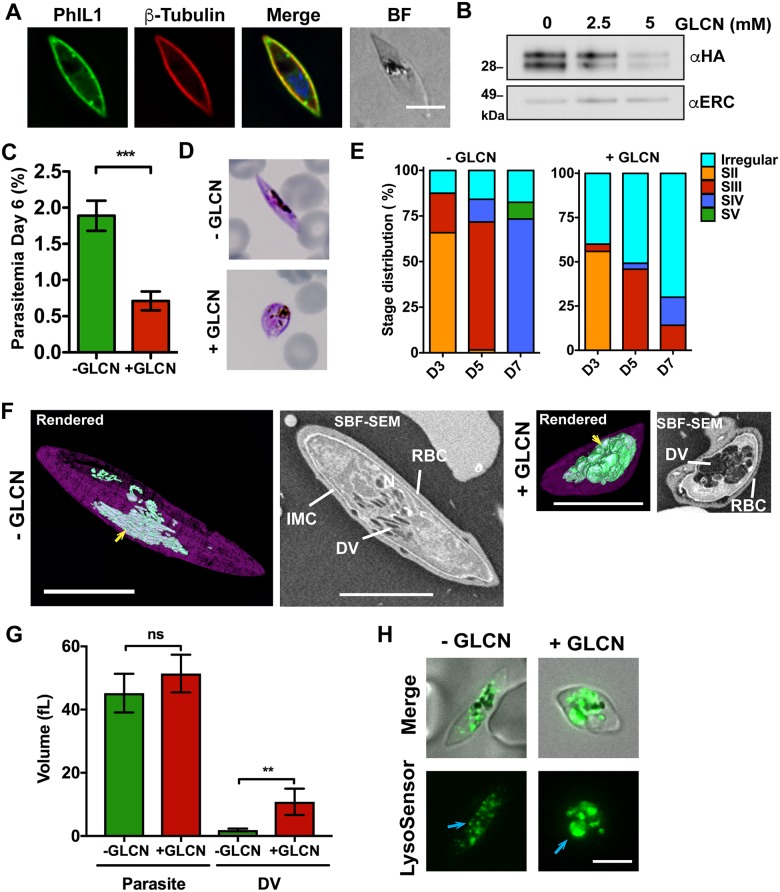
Fig 5. Knockdown of PhIL1 arrests gametocyte development.
(A) PhIL1-HA-glmS (green) is located at the parasite periphery, closely associated with β-tubulin (red). (B) Stage IV PhIL1-HA-glmS gametocytes treated with a range of glucosamine concentrations and analyzed by Western blotting. Probing with anti-HA shows efficient knockdown of the PhIL1-HA protein when compared to the anti-PfERC loading control. (C) Glucosamine-treated (+GLCN, 5 mM) and untreated (-GLCN, 0 mM) cultures were induced to form gametocytes and the parasitemia estimated at day 6. Data represent mean ± SEM; n = 3 experiments performed in triplicate. *** P <0.001, unpaired t-test. (D) Representative Giemsa-stained smears highlighting the altered morphology following PhIL1 knockdown. (E) Stage progression counts for PhIL1-HA-glmS parasites plus or minus glucosamine, highlighting the arrest in development following knockdown. Days 3, 5 and 7 only are shown. The complete data set, including wild type parasites plus or minus glucosamine, are presented in S6C and S6D Fig. Data from 2 separate experiments performed in triplicate. Mean values are shown. (F) SBF-SEM images of PhIL1-HA-glmS parasites, plus or minus glucosamine. Rendered models are shown on the left, highlighting the IMC in purple and the digestive vacuole in green. A swollen digestive vacuole (DV; yellow arrow) is observed in glucosamine-treated parasites. Scale bars: 5 μm. (G) Quantification of the SBF-SEM images. Mean volumes for the parasite and the digestive vacuole are shown. Data represent mean ± SEM. n = 5. ** P <0.01, unpaired t-test. (H) PhIL1-HA-glmS parasites plus or minus glucosamine were labeled with LysoSensor to highlight the acidic digestive compartments. Swollen compartments are seen in the treated group, but not in the untreated control group (blue arrows).

