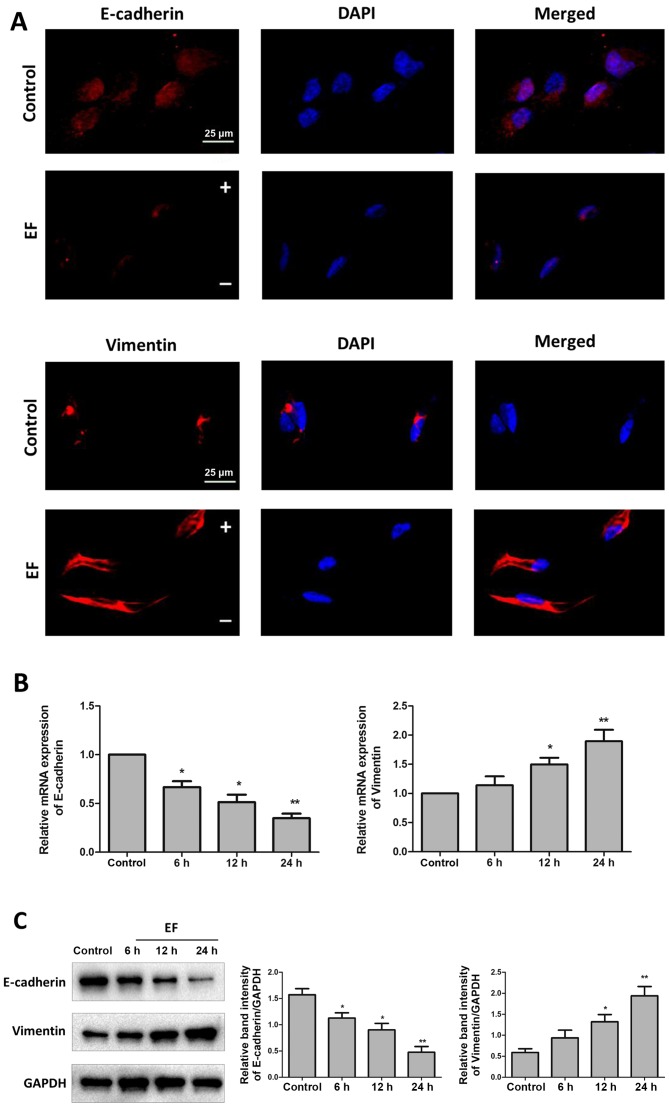
Figure 2.
EF exposure promotes expression of EMT markers in HLE-B3 cells. Cells were examined for expression of E-cadherin and Vimentin, without (control) or with (EF) exposure to an EF of 100 mV/mm. (A) Immunofluorescent staining of EMT markers (red) following 24 h EF exposure. Nuclei were counterstained with DAPI (blue). Scale bar, 25 µm. (B) Reverse transcription-quantitative polymerase chain reaction analysis following 6, 12, and 24 h EF exposure. (C) Western blot analysis following 6, 12, and 24 h EF exposure. Data were presented as mean + standard deviation. *P<0.05 and **P<0.01 vs. control. EF, electric field; EMT, epithelial-mesenchymal transition; -, cathode; +, anode.

