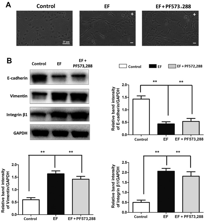
Figure 5.
Effect of FAK inhibition on EF-induced EMT. (A) Cell morphology was observed by microscopy in HLE-B3 cells following EF exposure, with or without pretreatment with the pFAK specific inhibitor PF-573,288. Scale bar, 25 µm. (B) Western blot analysis of HLE-B3 cells following EF exposure, with or without pretreatment with PF-573,288. Data were presented as mean + standard deviation. **P<0.01, with comparisons indicated by lines. FAK, focal adhesion kinase; EF, electric field; EMT, epithelial-mesenchymal transition; p, phosphorylated; -, cathode; +, anode.

