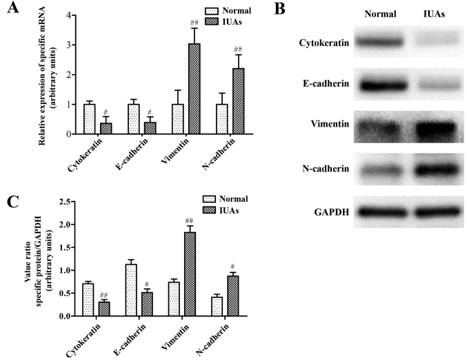
Figure 3.
Changes of epithelial mesenchymal transition-associated proteins in a murine IUAs model. (A) Epithelial marker proteins, cytokeratin and E-cadherin and the mesenchymal marker proteins, vimentin and N-cadherin, were measured by reverse transcription-quantitative polymerase chain reaction. (B) Representative images of western blot experiments. (C) Densitometry quantification of cytokeratin, E-cadherin, vimentin and N-cadherin by western blot analysis. #P<0.05, ##P<0.01 vs. normal group. IUAs, intrauterine adhesions.

