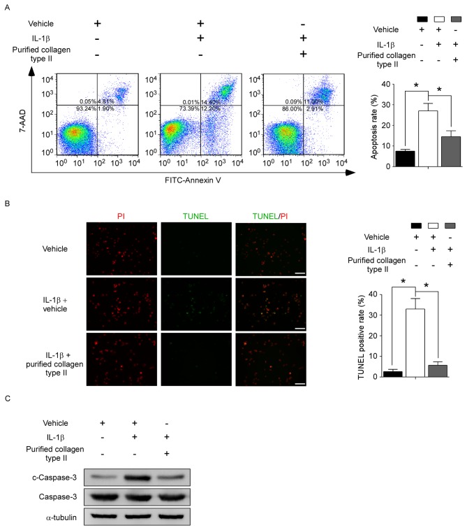
Figure 3.
Collagen type II protects human NP cells from IL-1β-induced apoptosis. Human NP cells were treated with or without vehicle (0.05 M acetic acid), 10 ng/ml IL-1β and/or 100 µg/ml purified collagen type II for 48 h, as indicated. (A) Cells were stained with FITC-conjugated Annexin V and 7-AAD and analyzed by flow cytometry. Representative plots and quantification are shown as means ± standard deviation of three independent experiments. (B) TUNEL assay was evaluated by fluorescence microscopy. Green fluorescence indicates apoptotic TUNEL-positive cells, while red fluorescence indicates PI-stained nuclei. Representative images and quantification are shown as means ± standard deviation of three independent experiments. Scale bar, 100 µm. (C) Protein levels of c-caspase-3 and total capase-3 were evaluated by western blotting. *P<0.05, with comparisons indicated by brackets. NP, nucleus pulposus; IL, interleukin; FITC, fluorescein isothiocyanate; 7-AAD, 7-aminoactinomycin D; TUNEL, terminal deoxynucleotidyl-transferase-mediated dUTP nick end labelling; PI, propidium iodide; c, cleaved.

