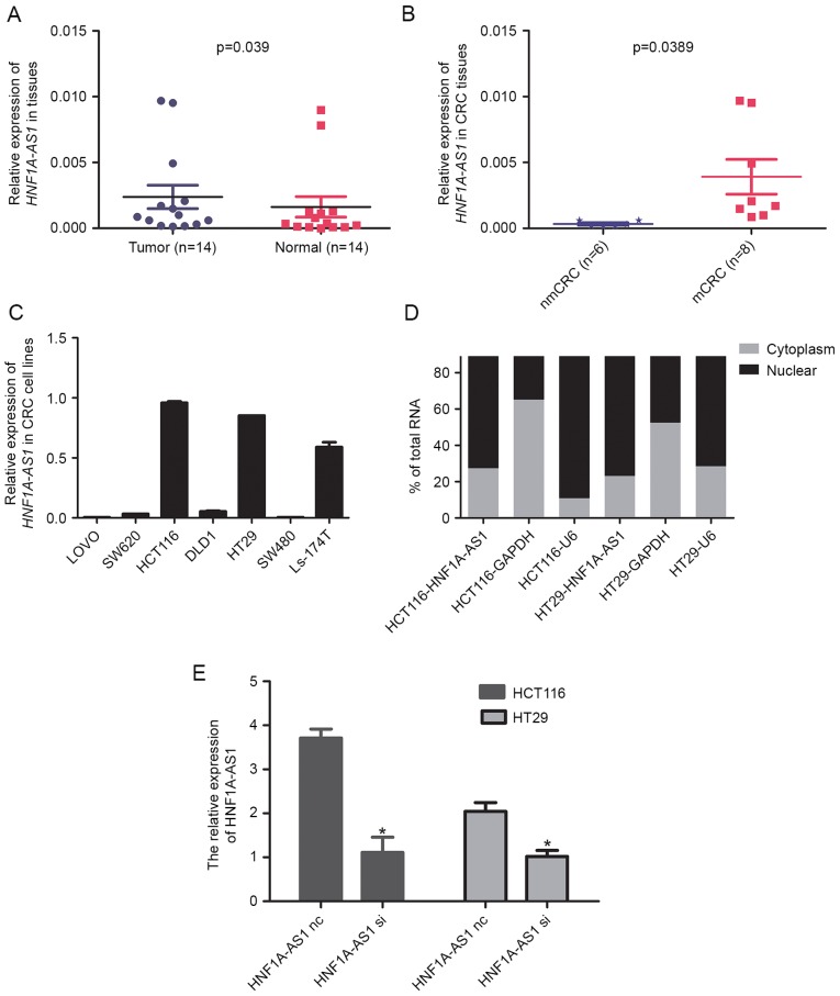
Figure 1.
Analysis of HNF1A-AS1 expression in CRC tissues and cells. (A) Relative expression of HNF1A-AS1 in paired CRC tissues and adjacent non-tumor tissues (n=14). HNF1A-AS1 expression was examined by RT-qPCR and normalized to GAPDH expression. (B) The HNF1A-AS1 expression in CRC tissues with (mCRC) or without (nmCRC) lymph-node metastasis. (C) Relative expression of HNF1A-AS1 in seven CRC cell lines. HNF1A-AS1 expression was examined by RT-qPCR and normalized to GAPDH expression. (D) HNF1A-AS1 cell localization, as identified using RT-qPCR in fractionated HCT116 and HT29 cells. GAPDH and U6 were used as cytosolic and nuclear fractionation indicators, respectively. (E) Relative expression of HNF1A-AS1 was measured in HCT116 and HT29 cells by RT-qPCR following transfection with either a specific siRNA (si) or a negative control siRNA (nc). *P<0.05 vs. nc group. HNF1A-AS1, HNF1A antisense RNA 1; CRC, colorectal carcinoma; RT-qPCR, reverse transcription-quantitative polymerase chain reaction; m, metastatic; nm, non-metastatic; U6, U6 small nuclear RNA; siRNA, small interfering RNA; nc, negative control.

