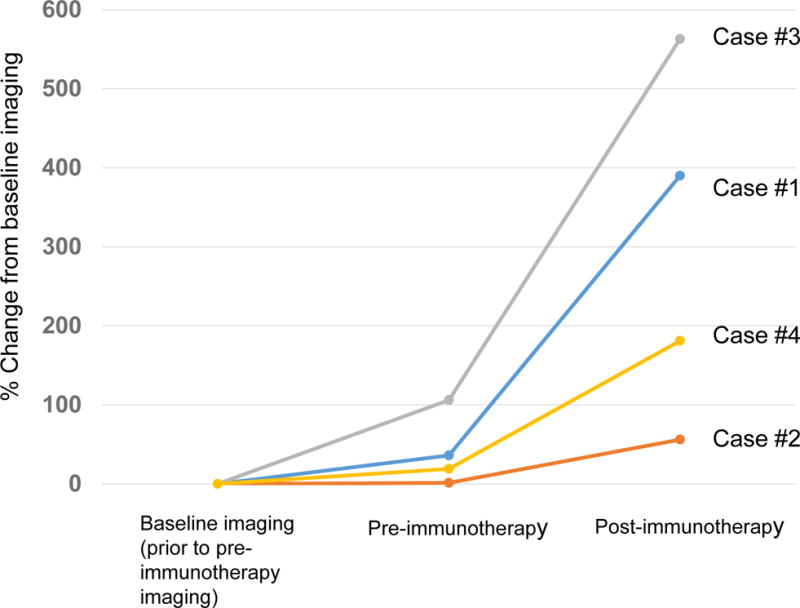
Figure 2.

Rate of change in growth pattern in four cases with MDM2 amplification that progressed rapidly while on immunotherapy. Rate of progression is compared from about 2 months prior to immunotherapy (baseline) to image immediately before immunotherapy (pre-immunotherapy), and then to first imaging after immunotherapy. Percent change was evaluated with immune-related response criteria (18).
Case #1: Pre-immunotherapy imaging showed ~36% increase in size of tumors when compared to baseline imaging. After immunotherapy, tumor progressed with 390% increase in lesions when compared to baseline imaging (258% increase from pre-immunotherapy) (7.2-fold increase in progression pace compared to ~2 months before immunotherapy). New liver masses also appeared.
Case #2: Pre-immunotherapy imaging showed 1.3% increase in size of tumors when compared to baseline imaging. After immunotherapy, tumor progressed with 56% increase when compared to baseline imaging (55% increase from pre-immunotherapy) (42.3-fold increase in pace of progression compared to the ~2 months before immunotherapy). New masses also appeared.
Case #3: Pre-immunotherapy imaging showed 106% increase in size of tumors when compared to baseline imaging. After immunotherapy, patient’s tumor progressed with 563% increase compared to baseline (242% increase compared to pre-immunotherapy) (~2.3 fold increase in rate of progression compared to the 2 months before immunotherapy). Multiple new large masses were seen.
Case #4: Pre-immunotherapy imaging showed a 19% increase in size of tumors when compared to baseline imaging. After immunotherapy, patient’s tumor progressed with 181% increase from baseline imaging (135% increase from pre-immunotherapy) (7.1-fold increase in progression pace compared to 2 months before therapy).
