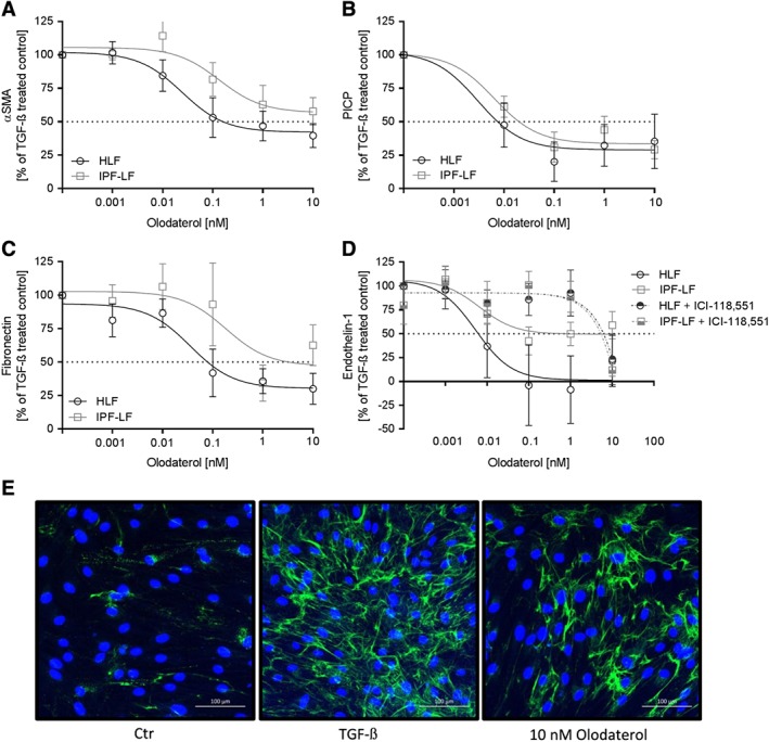
Figure 2.

Olodaterol attenuates TGF‐β‐stimulated protein expression of primary HLF. Fibroblasts from control donors (HLF) and patients with IPF (IPF‐LF) were pre‐incubated with different concentrations of olodaterol and subsequently stimulated with TGF‐β (4 ng·mL−1) for 48 h in the presence of the compound. α‐SMA protein expression was measured in cell lysates by an MSD Western replacement assay (A). Pro‐collagen I C‐peptide (B), fibronectin (C) and ET‐1 (D) expression was measured in supernatants by elisa. Basal levels of 16 ng·mL−1, 1.5 μg·mL−1 and 0.2 pg·mL−1 increased to 40 ng·mL−1, 2.5 μg·mL−1 and 5 pg·mL−1 respectively. Effect of olodaterol on ET‐1 protein expression in HLF and IPF‐LF in the presence of ICI‐118,551 (30 nM) (D). Data are expressed as normalized protein expression (100% is expression with TGF‐β stimulation). Data shown are means ± SEM of n = 5 different donors for HLF and n = 5 different donors for IPF cells. Horizontal dotted line is 50% inhibition of the TGF‐β‐induced effect. Representative image of TGF‐β‐induced collagen I assembly and inhibition by 10 nM olodaterol in HLF in a ‘scar‐in‐a‐jar’ assay (E). Unstimulated cells (Ctr) were compared to TGF‐β‐stimulated cells (TGFβ) and TGFβ‐stimulated and olodaterol‐treated cells (10 nM Olodaterol).
