Figure 6.

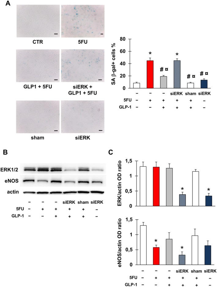
ERK 1/2 is implicated in 5FU toxicity on endothelial cells. (A) Representative images and percentage of EA.hy926 cells stained for SA β‐gal after the following treatments: no treatment (CTR); exposure to 5FU; pre‐incubation with GLP‐1 followed by exposure to 5FU; transfection with ERK1/2 siRNA (siERK), pre‐incubation with GLP‐1 and then exposure to 5FU; transfection with sham siRNA (sham); transfection with siERK. Magnification of pictures is 200×, and bars correspond to 50 μm. (B, C) Representative western blots for ERK1/2 and eNOS (B) and optical density (OD) of the protein bands normalized to that of actin (C) after the treatments described in (A). Graphs show mean ± SEM of five independent experiments. Comparisons were made by means of ANOVA followed by post hoc Tukey's multiple comparisons test. * Statistically significant versus CTR, # statistically significant versus 5FU and ¤ statistically significant versus siERK + GLP‐1 + FU.
