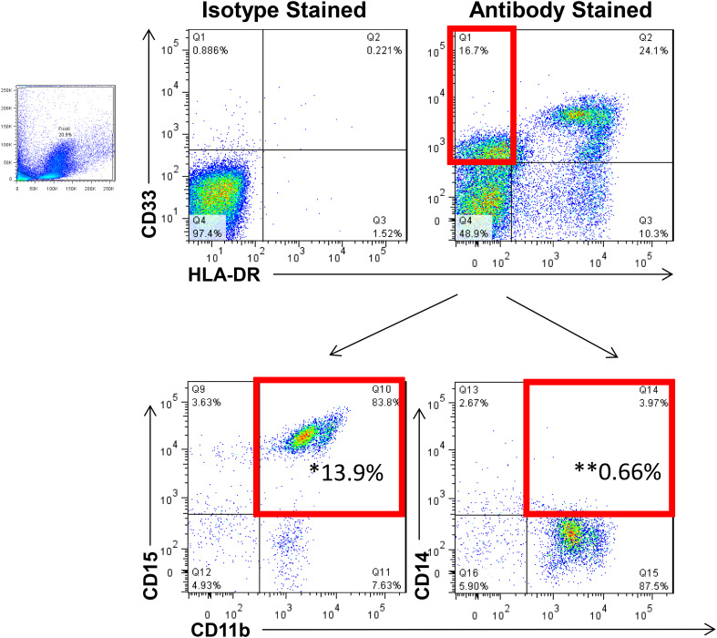
Fig. 1.
Representative flow cytometry staining for myeloid-derived suppressor cells. To quantify MDSC levels, flow cytometry was performed on cryopreserved PBMC from each patient. MDSC were identified as HLA-DR−, CD11b+, CD33+ cells with granulocytic and monocytic subsets expressing CD15 and CD14, respectively. *Granulocytic MDSC; **Monocytic MDSC. Data are representative of 24 patients analyzed at four time points each

