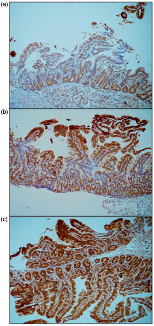
Figure 2.

Immunohistochemical staining intensity scoring. Three different specimens of normal duodenum, each stained with immunohistochemistry antibody for Uroguanylin, were scored by the blinded pathologist. Base magnification is 10× with an ocular of 10×. These slides demonstrate the scoring of (a) “1,” (b) “2,” and (c) “3” based on the overall intensity of the brown pigment in the intestinal epithelium and lamina propria.
