Figure 2.

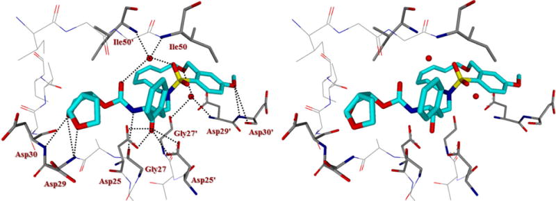
Stereoview of the X-ray structure of inhibitor 5j (turquoise)-bound HIV-1 protease (PDB code: 5WLO). All strong active site hydrogen bonding interactions of inhibitor 5j with HIV-1 protease are shown as dotted lines.

Stereoview of the X-ray structure of inhibitor 5j (turquoise)-bound HIV-1 protease (PDB code: 5WLO). All strong active site hydrogen bonding interactions of inhibitor 5j with HIV-1 protease are shown as dotted lines.