
Fig. 6.
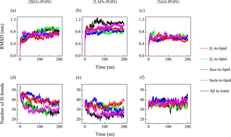
Structural stability analysis of the three protofibrillar Aβ9-40 trimers in the presence of POPG lipid bilayers. For each Aβ-POPG system (2BEG-POPG, 2LMN-POPG, 2M4J-POPG), we show the time evolution of the Cα-RMSD (a, b, c) and the number of backbone hydrogen bonds (d, e, f) of Aβ trimer in MD runs starting from four states: β1-to-lipid (red), β2-to-lipid (green), face-to-lipid (blue) and back-to-lipid (pink). For comparison, the results for each Aβ trimer in water without POPG bilayers are also given (black).