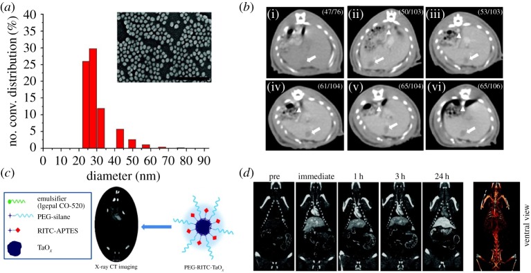
Figure 2.
(a) Hydrodynamic size distribution and scanning electron microscopy (SEM) image of PEG-coated gold nanoparticles. Scale bar, 300 nm. (b) In vivo CT serial imaging of a rat hepatoma model before (i) and 5 min (ii), 1 h (iii), 2 h (iv), 4 h (v), 12 h (vi) after intravenous administration of PEG-coated gold nanoparticles. Arrows indicate the hepatoma regions, and arrowheads indicate the aorta. (c) Schematic of RITC (rhodamine isothiocyanate)-doped tantalum oxide nanoparticles for X-ray CT imaging. (d) In vivo CT serial imaging of a rat after intravenous injection of RITC-doped tantalum oxide nanoparticles. Reproduced with permission from [41] and [10].

